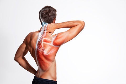
The neck, also called the cervical spine, is a well-engineered structure of bones, nerves, muscles, ligaments, and tendons. The cervical spine is delicate—housing the spinal cord that sends messages from the brain to control all aspects of the body—while also remarkably strong and flexible, allowing the neck to move in all directions.
Anatomy of the Cervical Spine
Anatomy of the Cervical Spine
Anatomy of the Cervical Spine
The cervical spine has 7 stacked bones called vertebrae, labeled C1 through C7. The top of the cervical spine connects to the skull, and the bottom connects to the upper back at about shoulder level. As viewed from the side, the cervical spine forms a lordotic curve by gently curving toward the front of the body and then back. This complex structure includes 7 small vertebrae, intervertebral discs to absorb shock, joints, the spinal cord, 8 nerve roots, vascular elements, 32 muscles, and ligaments.
The nerve roots stem from the spinal cord like tree branches through foramen in the vertebrae. Each nerve root transmits signals (nerve impulses) to and from the brain, shoulders, arms, and chest.
A vascular system of 4 arteries and veins run through the neck to circulate blood between the brain and the heart. Joints, muscles, and ligaments facilitate movement and serve to stabilize the structure. Neck mobility is matchless. It is capable of moving the head in many directions: 90° of flexion (forward motion), 90° of extension (backward motion), 180° of rotation (side to side), and almost 120° of tilt to either shoulder.
Meet our Doctors

Dr. Santoshi Kurada
MBBS, MD, Fellow in Critical Care and Pain Medicine (UK)

Dr. Swagatesh Bastia
MBBS, MS (Ortho), Fellow in Trauma and Orthopaedics (UK), Fellow in Pain Management

Dr. Faraz Ahmed Syed
MBBS, MS (Orthopaedics), AO Spine Surgery Fellow (UK)

Dr. Wiquar Ahmed
MBBS, DA, FIPM, CIPS

Dr Roshan Adappa
MBBS, MD (Anaesthesiology), FIASP, MMed -Pain Management (UK), European Diploma in Pain Medicine (EDPM, Belgium)
Dr. Shubha V Hegde
MBBS, MD, DNB(Anaesthesia), EDPM-1 (Europe), FIAPM, FIPM, CCEPC

Dr. Pramodh
MBBS,DFM,CCEBDM,MLSM
© 2020-21 Alleviatepainclinic. All rights reserved.